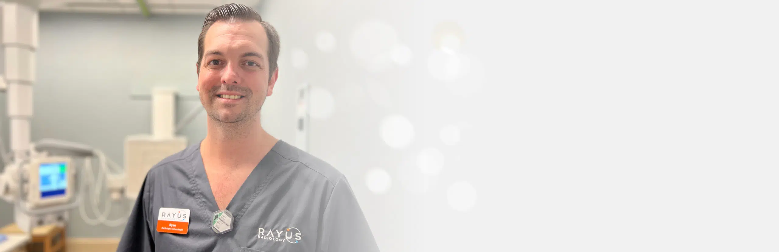
Ultrasound
Ultrasound imaging, also called ultrasound scanning or sonography, is a method of obtaining images from inside the human body through the use of high frequency sound waves. The reflected sound wave echoes are recorded and displayed as a real-time visual image. No ionizing radiation (x-ray) is involved in ultrasound imaging. Obstetric ultrasound refers to the specialized use of sound waves to visualize and thus determine the condition of a pregnant woman and her embryo or fetus.
Ultrasound is a useful way of examining many of the body's internal organs, including the heart, liver, gallbladder, spleen, pancreas, kidneys, and bladder. Because ultrasound images are captured in real-time, they can show movement of internal tissues and organs, and enable physicians to see blood flow and heart valve functions. This can help to diagnose a variety of heart conditions and to assess damage after a heart attack or other illness.
Ultrasound Exam Preparations
|
 |

Ultrasound Images
 |
Lung Ultrasound |
 |
OB Ultrasound |
 |
Pancreas Ultrasound |

Ultrasound Services Offered
Ultrasound
- Abdomen
- Pelvis
- Transvaginal
- OB
- Carotid
- Aorta
- Thyroid
- Renal
- Bladder
- Scrotum/testicles
- Venous Ultrasound
- Arterial
- Echo

Ultrasound Exam Preparations
Carotid, Thyroid, Breast, Scrotal, Echocardiography: No preparation is required.
Gallbladder, Liver, Abdominal, Pancreas: No food for 6 hours prior to the exam.
Pelvis / Bladder Ultrasound: Drink & Finish 32 oz of clear liquids 1 hour prior to exam. Do not urinate.
Make An Appointment
Diagnostic Center of America Home
|